|
Deformability based cell margination—A simple microfluidic design for
malaria-infected erythrocyte separation
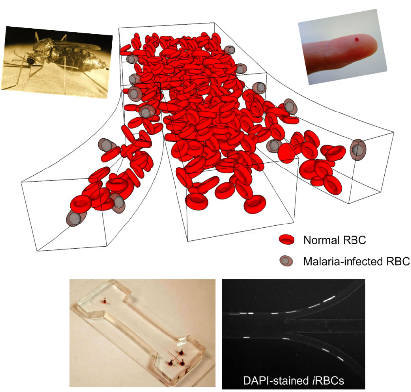
In one of their latest publications in the journal "Lab on a Chip" (H.W. Hou, A.A.S. Bhagat, A.G.L. Chong, P. Mao P, K.S.W. Tan, J. Han and C.T. Lim, "Deformability based cell margination – A simple microfluidic design for high throughput malaria infected erythrocyte separation", Lab on a Chip, vol. 10, 2605 (2010)), BioSyM and NUS researchers describe a deformability-based separation method for iRBCs separation in a microfluidic device, inspired by the in vivo phenomenon of leukocyte margination.
In blood vessels with luminal diameter less than 300mm, red blood cells (RBCs) which are smaller in size and more deformable than leukocytes, migrate to the axial centre of the vessel due to flow velocity gradient within the vessels. This phenomenon displaces the leukocytes to the vessel wall and is aptly termed as margination. This work demonstrate using microfluidics that stiffer malaria-infected RBCs (iRBCs) behave similar to leukocytes and undergo margination towards the sidewalls. This provides better understanding of the hemodynamic effects of iRBCs in microcirculation and its contribution to pathophysiological outcome relating to cytoadherence to endothelium. In this work, cell margination is mimicked for the separation of iRBCs from whole blood based on their reduced deformability. The malaria infected sample was tested in a simple long straight channel microfluidic device fabricated in polydimethylsiloxane. In this microchannel, cell margination was directed along the channel width with the iRBCs aligning near each sidewall and then subsequently removed using a 3-outlet system, thus achieving separation. Tests were conducted using ring stage and late trophozoite/schizont stage iRBCs. Device performance was quantified by analyzing the distribution of these iRBCs across the microchannel width at the outlet and also conducting flow cytometry analysis. Results indicate recovery of ~75% for early stage iRBCs and >90% for late stage iRBCs at the side outlets. The simple and passive system operation makes this technique ideal for on-site iRBCs enrichment in resourcelimited settings, and can be applied to other blood cell diseases, e.g. sickle cell anemia and leukemia, characterized by changes in cell stiffness.
|
 |